In this chapter, we will discuss the biology of human hair and the surgical management of hair loss.
Hair is a biomaterial consisting of keratin. It is found abundantly over the biggest organ in the body, namely the skin. The hair transplant surgeon must know the anatomy of the hair follicle and understand the hair growth cycle to help plan and perform the procedure.
2.1. Anatomy of the hair follicle
Anatomically, hair is broadly divided into an upper, middle and lower segment. The upper segment contains the infundibulum and isthmus, the middle segment contains the bulge which stores stem cells and the inferior segment contains the bulb (Figure 20.1) (Schneider et al., 2009).
The hair follicle is a part of the epidermis that extends down into the dermis. The hair follicular unit (FU) is the grouping of terminal hairs, which can be seen under a microscope and contains approximately one to four hairs (Bernstein et al., 1998).
The infundibulum is the superficial part of the follicle. The isthmus is the part between the sebaceous gland duct opening and the bulge. The bulb is the deepest portion of the follicle and houses the stem cells that form matrix cells. The matrix cells keratinise to form the hair cortex and divide to form the remaining hair layers (Kahle et al., 1993).
The bulb also surrounds the dermal papilla, a structure which is responsible for the embryonic generation of a hair follicle and, in the fully formed follicle, contains capillaries, nerves and melanocytes (Blume-Peytavi et al., 2008).
The follicle is surrounded by many layers. From innermost to outermost are the layers of the hair shaft which can be seen above the skin: the medulla, cortex and outer cuticle. In the dermis, this is further enveloped by the inner root sheath, outer root sheath and connective tissue sheath (Figure 20.1).
2.1.1. Medulla
The medulla is in the centre of the shaft. It is sometimes absent in hair; when present (usually in coarse hair), it is often discontinuous along the length of hair (Krstic, 1991).
2.1.2. Cortex
The cortex represents 90% of the total weight of the shaft. It is made of spindle-shaped cortical cells filled with keratin. Young cortical cells contain melanin granules which determine the hair colour. These cells become elongated as they progress from the matrix. They are held within intercellular cement composed of keratin and lipids (Krstic, 1991; Kahle et al., 1993).
2.1.3. Cuticle
The cuticle is the outermost layer of the shaft and is made of keratin. It protects the inside of the hair shaft from damage. The cells forming the cuticle are also held within intercellular cement which is rich in lipids. The cells overlap one another and are said to resemble the slates on a roof (Krstic, 1991).
2.1.4. Inner root sheath
The inner root sheath extends from the isthmus to the base of the bulb. It consists of three layers: an inner cuticle, the Huxley layer and the outer Henle layer. The cells in these layers are identifiable by the presence of a structural protein called trichohyalin, which is a marker for hair follicular differentiation (Kahle et al., 1993).
2.1.5. Outer root sheath
The outer root sheath is found at the periphery and continues within the epidermis. It contains Golgi complexes, mitochondria and glycogen. A ‘bulge’ can be seen on the outer sheath where the erector pili muscle inserts. This bulge sits below the sebaceous gland and causes the gland to secrete sebum when it contracts (Kahle et al., 1993)
2.2. Embryology of the hair
Each hair on the body grows from a hair follicle. The induction and formation of an embryonic hair follicle is regulated by mesenchymal–epithelial interactions between dermal cells and epidermal stem cells. The primary hair germ begins as an epithelial bud. This bud projects down into the dermis to form the base of the follicle at week 12 (Figure 20.2). At week 14, the base of this follicle expands to form the hair bulb and is invaginated by mesoderm called the dermal papilla. By the 16th week of intrauterine life, the primary hair germ has grown deeper into the dermis and differentiates into many components of the primary hair follicle.
By the 18th week of intrauterine life, the hair emerges through the skin surface and by week 22 all of the hair follicles are formed. At this stage of life, there are approximately 5 million hair follicles on the body and 100,000 on the head (Dudek, 2010).
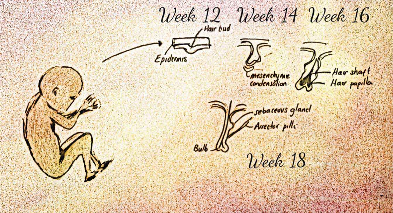
Figure 20.2. – Embryology of hair.
2.3. Hair life cycle
The lower portions of the hair follicles are involved in a lifelong growth cycle; the isthmus and infundibulum, however, remain stable. The cycle is characterised by periods of growth (anagen), periods of transformation (catagen) and rest periods (telogen) (Figure 20.3). These three separate periods of hair growth are not synchronous and occur independently of one another, which is important to avoid periods of extreme hair growth or loss (Price, 1999).
2.3.1. Anagen
During the anagen phase, the matrix of the hair follicle undergoes increased mitotic activity. Most scalp hair follicles are in the anagen phase for 90% of the hair life cycle; this phase lasts for 2–7 years. The rate of scalp hair growth is approximately 0.3 mm per day (Price, 1999).
2.3.2. Catagen
Approximately 1% of scalp hair is in this phase, which lasts for 2–3 weeks. The mitotic activity of the hair follicle decreases during this period and hair production diminishes. The deepest part of the hair follicle migrates up toward the isthmus (Price, 1999).
2.3.3. Telogen
Up to 10% of scalp follicles are in this phase, which lasts 2–3 months. It is characterised by the presence of club hair, i.e. fully keratinised hair that is ready for shedding from the hair follicle. Up to 100 telogen hairs are shed per day (Paus and Cotsarelis, 1999).
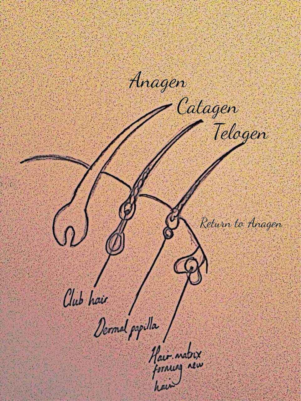
Figure 20.3. – The human hair growth cycle.